
In the evolving field of metabolic regulation and cellular bioenergetics, mitochondrial-derived peptides (MDPs) represent an important class of signaling molecules. Unlike nuclear-encoded signaling proteins, these specialized micro-proteins act as retrograde messengers that communicate cellular stress directly to the nucleus. To analyze these metabolic regulatory vectors in preclinical models, research teams investigating metabolic flexibility are prioritizing MOTS-c peptide Canada protocols to quantify how mitochondrial signaling influences system-wide homeostasis.
This technical guide examines the structural composition of MOTS-c, details its metabolic pathways, outlines standardized reconstitution procedures for 10mg laboratory vials, and provides analytical quality controls necessary for validating experimental data.
MOTS-c Structural Architecture & Nuclear Translocation
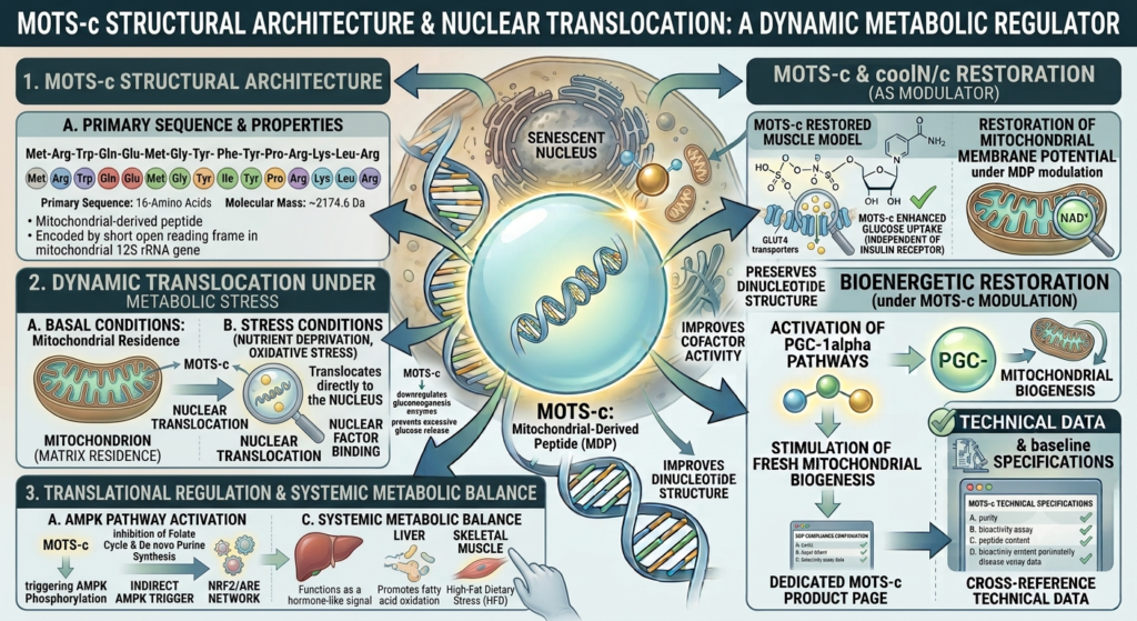
MOTS-c is a 16-amino acid mitochondrial-derived peptide encoded by a short open reading frame within the mitochondrial 12S ribosomal RNA gene. Its defined primary sequence is Met-Arg-Trp-Gln-Glu-Met-Gly-Tyr-Ile-Phe-Tyr-Pro-Arg-Lys-Leu-Arg, yielding a molecular mass of approximately 2174.6 Da.
In cellular assays, investigating mitochondrial peptide research frameworks demonstrates that MOTS-c behaves dynamically under metabolic stress. Under basal conditions, the peptide resides within the mitochondrial matrix; however, upon nutrient deprivation or oxidative stress, it translocates directly to the nucleus to regulate transcriptional networks:
- AMPK Pathway Activation: It indirectly triggers AMP-activated protein kinase (AMPK) phosphorylation by inhibiting the folate cycle and de novo purine synthesis, elevating intracellular AICAR levels.
- Nuclear Factor Binding: Upon nuclear translocation, it interacts with transcription factors like NRF2 to orchestrate antioxidant response element (ARE) gene expression.
- Systemic Metabolic Balance: It functions as a hormone-like signal, promoting fatty acid oxidation and glucose clearance in skeletal muscle models under high-fat dietary stress.
Insulin Sensitivity Dynamics & Glucose Homeostasis
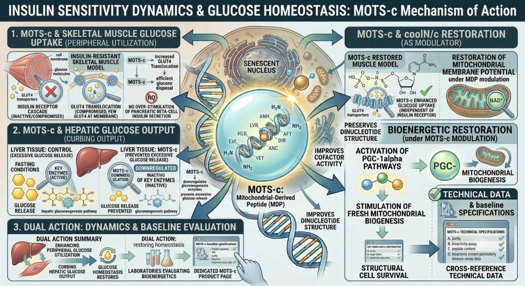
A major target in any comprehensive MOTS-c insulin sensitivity study is quantifying how the peptide enhances glucose uptake independently of standard insulin receptor cascades. In insulin-resistant skeletal muscle models, MOTS-c administration increases GLUT4 transporter translocation to the cell membrane, restoring efficient glucose disposal without over-stimulating pancreatic beta-cell insulin secretion.
Furthermore, MOTS-c downregulates key enzymes involved in hepatic gluconeogenesis, preventing excessive glucose release from liver tissue under fasting conditions. This dual action—enhancing peripheral glucose utilization while curbing hepatic glucose output—positions MOTS-c as a key subject in metabolic disease models. Laboratories evaluating these bioenergetics cross-reference baseline specifications against technical data on the dedicated MOTS-c product page.
Reconstitution & Volumetric Calculations for MOTS-c 10mg Vials
To preserve tight experimental control and ensure data reproducibility across repetitive assay models, meticulous preparation protocols are critical. Subtle shifts in target concentrations can introduce unwanted baseline drift in cellular responses, meaning that establishing a precise, standardized reconstitution map is a mandatory prerequisite for any formal study design.
Lyophilized chemical cakes must be carefully brought to a liquid state using an appropriate sterile diluent. For extended observational studies, the industry standard is Bacteriostatic Water (0.9% Benzyl Alcohol), which provides an effective anti-microbial barrier to inhibit bacterial growth over multi-week testing cycles. The table below details standard dilution configurations across common laboratory 10mg vial structures:
| Vial Mass Total | Reconstitution Liquid Volume | Resulting Core Concentration | Standard Micro-Dose Aliquot |
|---|---|---|---|
| 20 mg (20,000 mcg) | 1.0 mL | 20,000 mcg / mL (20 mg/mL) | 2,000 mcg (2 mg) per 0.10 mL unit |
| 20 mg (20,000 mcg) | 2.0 mL | 10,000 mcg / mL (10 mg/mL) | 1,000 mcg (1 mg) per 0.10 mL unit |
| 20 mg (20,000 mcg) | 2.5 mL | 8,000 mcg / mL (8 mg/mL) | 800 mcg (0.8 mg) per 0.10 mL unit |
Analytical Purity Verification & Sourcing Controls
When domestic research bodies arrange to procure chemical compounds for metabolic assays, validating raw material purity represents the primary safeguard against compromised experimental readouts. Minor faults during solid-phase peptide synthesis can yield truncated amino acid sequences or residual trifluoroacetic acid (TFA) salts, which can induce unexpected cell toxicity or alter metabolic responses in live assays.
Every genuine batch of MOTS-c must be validated by independent testing using High-Performance Liquid Chromatography (HPLC) to confirm a chemical purity rating exceeding 98.0%. Simultaneously, Mass Spectrometry (MS) analysis should be used to confirm that the observed mass matches the theoretical profile of 2174.6 Da. Vials that reveal signs of early moisture ingress—such as a sticky, clumped, or discolored appearance of the dry powder cake—should be discarded immediately, as unchecked moisture initiates rapid hydrolysis and breaks down the peptide chain.
Handling Protocol & Mitochondrial Peptide Stability
As a short mitochondrial-derived peptide, MOTS-c requires precise cold-chain management to protect its primary amino acid sequence from mechanical shearing and ambient degradation:
- Lyophilized Powder Maintenance: Sealed, un-reconstituted MOTS-c vials should remain in deep-freeze storage at -20°C, shielded from light exposure. Sub-zero temperatures isolate the crystalline structure from thermal stress prior to reconstitution.
- Reconstitution Protocol: Gently trickle sterile diluent along the glass wall into the cake. Gently roll the vial between your palms until the powder is entirely dissolved. Never agitate violently, as shear forces can alter peptide folding.
- Refrigerated Storage Parameters: Keep the reconstituted liquid continuously refrigerated at 2°C to 8°C. Reconstituted MOTS-c should be fully utilized within 30 days to prevent chemical degradation in liquid media.
Summary: Horizons in Mitochondrial Signaling Research
Representing a distinct class of mitochondrial-derived peptides, MOTS-c acts as an active metabolic regulator capable of translocating to the nucleus during cellular stress. In vitro and in vivo models confirm its role in activating the AMPK pathway, improving skeletal muscle insulin sensitivity, and maintaining systemic metabolic homeostasis. To yield reliable, peer-review-quality data in mitochondrial signaling trials, research teams should ensure strict adherence to cold-chain storage at -20°C and precise reconstitution using sterile Bacteriostatic Water.
Metabolic Support
$74.99
Longevity & Vitality
$89.99

References
1. Lee, C., et al. (2015). The mitochondrial-derived peptide MOTS-c promotes metabolic homeostasis and reduces obesity and insulin resistance. Cell Metabolism, 21(3), 443-454. https://doi.org/10.1016/j.cmet.2015.02.009
2. Kim, K. H., et al. (2018). The mitochondrial-encoded peptide MOTS-c translocates to the nucleus to regulate nuclear gene expression in response to metabolic stress. Cell Metabolism, 28(3), 516-524. https://doi.org/10.1016/j.cmet.2018.06.008
3. Reynolds, J. C., et al. (2021). MOTS-c is an exercise-induced mitochondrial-encoded regulator of physiology and healthspan. Nature Communications, 12(1), 320. https://doi.org/10.1038/s41467-020-20790-0